

Skin Tumours in Horses
by Dr Melissa Bain
Tumours found in the skin of horses are relatively common and are a sign of cancer/neoplasia. In horses there are 3 main types of skin tumours that we find which have differing treatment and prognosis – these are squamous cell carcinomas, sarcoids, and melanomas. Tumours can be benign and remain localised to that region, or they can be malignant and expand into surrounding tissues or distant sites. Early recognition, identification, and treatment of these tumours is crucial for the best outcome for the horse and owner.
How do we recognise a possible skin tumour?
Skin tumours have a wide range of appearances, so we must be suspicious of a skin cancer whenever there is a significant change to the appearance of the skin. Some possible presentations of cancers include;
- Flat, hairless areas
- Smooth, firm lumps (haired and unhaired)
- Wart-like masses
- Fleshy, proud-flesh like masses
- Non-healing wounds

If you would like to book a skin consult please fill in the form below.
How can we identify what type of cancer it is?
Some tumours can be very typical of their type such as black-pigmented masses on a grey horse are typically melanomas, however sometimes we cannot tell immediately what type it is. This is particularly true with sarcoids which have a huge range of appearances. Next steps for identification may include a biopsy where we take part or the whole mass off surgically and send it for histopathology where they can identify the cell types present and which cancer it is definitively. If we suspect a sarcoid, we work in close conjunction with Professor Knottenbelt (Equine Medical Solutions UK) who is the leading expert in sarcoid identification and treatment, submitting photos/history to the oncologists for ongoing treatment and management plans.
What causes skin cancers?
Skin cancers have a wide range of causes. Some may be related to UV or chemical exposure to the skin, such as mucosal squamous cell carcinomas on pink-skinned horses. Some have a genetic component such as those melanomas seen on grey horses. We do not yet fully understand the cause of sarcoids but there is evidence of a genetic and viral component, with localised spread on a horse occurring via flies.
What are the treatment options for skin cancer?
Treatment strategies for these cancers will depend on many factors including the type of cancer, malignancy, location of the cancer on the body, age of the horse, and the financial budget. Strategies may be singular but we can also use multiple treatment options together for the best result. These may include;
- Topical chemotherapy (AW cream) or injectable chemotherapy
- Surgical excision or debulking of masses
- Cryotherapy using liquid nitrogen
- C02 laser surgery


Squamous Cell Carcinomas
What are the common sites for SCC?
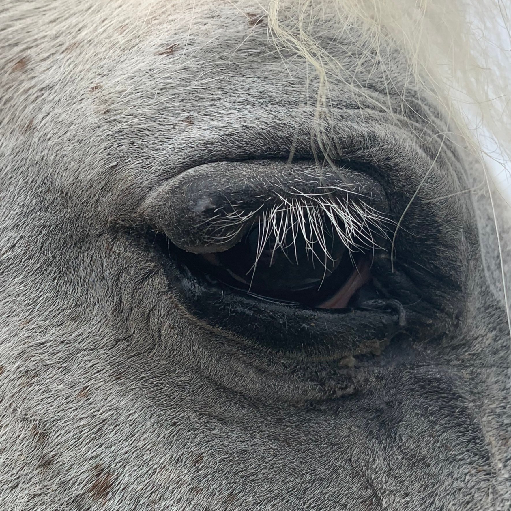
Squamous cell carcinoma (SCC) is a common skin cancer and occurs where there are squamous cells which includes the skin, penis, vulva, mouth, nasal cavity, and stomach. The most common site in horses is typically the penis, which is why it’s always recommended to closely check and clean the sheath/penis annually. Other common sites are the nose and the eyes (orbit and 3rd eyelid).
What causes SCC?
The cause of SCC has been linked to UV damage with increased risk in areas that are prone to sunburn (nose and eyes), and it has also linked to carcinogens in smegma (sebum, dead cells, dirt) on the penis and clitoral fossa.
How do we treat SCC?
SCC is malignant and is typically aggressive, spreading into surrounding tissue or increasing in size rapidly, so early identification and treatment whilst lesions are small offers the best prognosis for recovery. If the tumour is small and the margins (i.e. size of the tumour) are clear, then surgical excision is the treatment of choice. For small tumours we can use cryotherapy under sedation to kill the tumour cells by causing them to freeze. Cryotherapy can also be used to debulk/reduce-size of tumours before further treatment. Referral for CO2 laser surgery is also commonly used. We may use injectable or topical chemotherapeutic agents in conjunction with surgical excision.
What is the prognosis of SCC?
With small lesions that can be removed with cryotherapy or surgically excised with good margins, the prognosis of a full recovery is good. With extensive or aggressive SCC the treatment options may be limited, for example with extensive penile SCC the only option may be to perform a partial amputation.

If you would like to book a skin consult please fill in the form below.


Melanomas
What horses are affected by melanomas?
Melanomas are most commonly found in grey horses; >80% of grey horses will have one or melanomas in their lifetime. This rate increases with certain breeds such as Andalusians, Arabs, and Percherons. They can develop at any age but are more common in horses >10years. Typically they are benign, but they have the potential to become malignant at any time. However, melanomas can also be found in non-grey horses, and these tend to be more dangerous with a higher rate of malignancy.
What are the common sites for melanomas?
Melanomas can be found anywhere on skin, but the most common site is the skin of the perineum (around anus and base of tail). They can also be found around/in eyes, mouth/lips, penis, parotid salivary glands, and lymph nodes. They may also be found internally from the spread of a malignant melanoma in the skin.
What causes melanomas?
Unlike humans, melanomas in grey horses are not linked to UV exposure, and it is more likely caused by a genetic susceptibility by the grey colour genes +/- breed predisposition. However, melanomas in non-grey horses are often triggered by a mutation caused by UV or another carcinogen.
How/when do we treat melanomas?
We recommend the use of a “melanoma map” for all grey horses to measure and monitor the size and location of melanomas on your horse. Treatment of melanomas is typically through surgical excision +/- injectable chemotherapy at the time of surgery, and is best suited to small, individual tumours. Whether we decide to treat melanomas is dependent on many different factors some of which include;
- Type – melanomas on non-grey horses are typically treated
- Location – is it comprising bodily function or interfering with tack
- Malignancy – if no longer benign and growing in size
What is the prognosis of melanomas?
For the vast majority of grey horses, melanomas remain benign and do not negatively impact their quality of life and do not affect their lifespan. However, melanomas that become malignant and grow rapidly offer a poorer prognosis. The location of these is also important – those in the head can majorly affect nerve function and cause respiratory issues, and large perineal masses can cause defecation and urination issues. If the tumours ulcerate they can become infected and painful which offers a very poor prognosis. Additionally, those melanomas left untreated in non-grey horses offer a guarded prognosis.

Sarcoids
What are sarcoids?
Sarcoids are a type of skin cancer – they are tumours of fibroblast which are cells found in the skin and underlying tissues. They are very common and can affect horses of any age but are more common in middle-aged or older horses. We are still continuing to learn why sarcoid occur but there is evidence of a genetic and viral component, with localised spread on a horse occurring via flies.
Where are common sites for sarcoids?
Sarcoids can occur anywhere on the skin but are most commonly found at sites where flies may feed including the face, eyes, legs, and groin region. They can remain static for many months or even years, but growth is often triggered from trauma or injury to the area where they have been growing, causing an acceleration of growth potentially becoming a very aggressive lesion. Non-healing wounds may be due to sarcoid formation.
What do we do if we suspect a sarcoid?
We work in close conjunction with Professor Knottenbelt (Equine Medical Solutions UK) who is the leading expert in sarcoid identification and treatment, submitting photos/history to the oncologists for ongoing treatment and management plans. When we treat sarcoids can be a difficult decision, as effective treatment involves a significant time and cost commitment, and prognosis can be highly variable dependent on the type and location of the sarcoid(s).
How do we diagnose sarcoids?
Sarcoids can often be visually diagnosed by a veterinarian, however sometimes we are not able to differentiate sarcoids from other skin lesions or other skin cancers easily. If we are committed to treating the potential sarcoid we may take a biopsy to confirm, but as biopsy causes trauma to the sarcoid which can trigger further growth, it is not recommended to biopsy a lesion if there is no intention of treating it.
What are the 6 types of sarcoids?
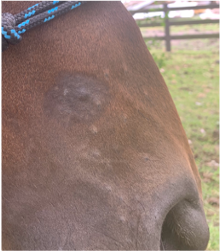
1. Occult – early stage, circular hairless area with thickened skin +/- small nodules
2. Verrucose – wart like lesions often with surrounding occult “halo”
3. Nodular – firm nodules under skin that may ulcerate
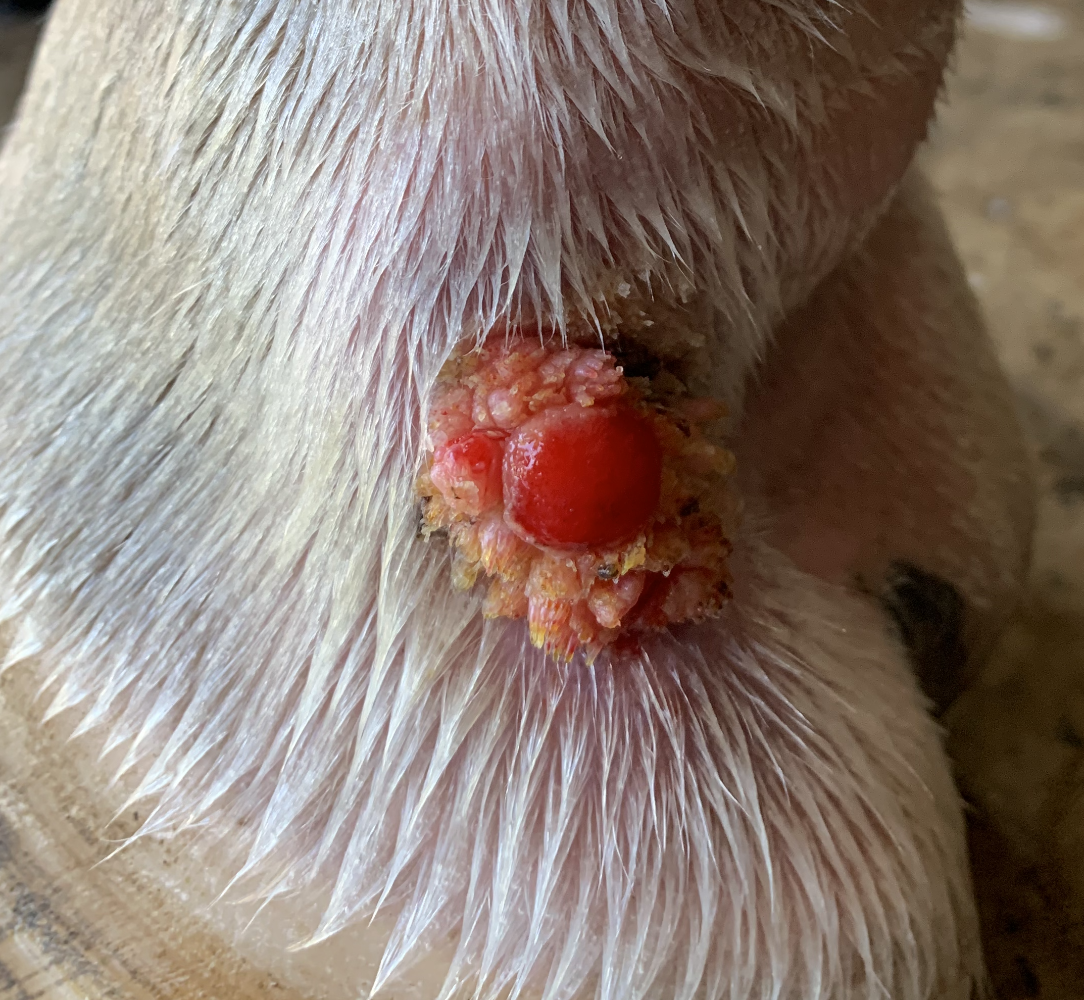
4. Fibroblastic – fleshy, highly vascular, often occur after injury to prior sarcoid
5. Mixed – can be a combination of sarcoid types at one location
6. Malignant – highly aggressive nodular and fibroblastic lesions that ulcerate
How do we treat sarcoids?
We typically treat with a topical chemotherapeutic agent (AW cream) obtained from the UK in consultation with Prof Knottenbelt that is applied by a veterinarian on a set protocol. Surgical excision of sarcoids has a varying success dependent on the type of sarcoid, and can be used in conjunction with injectable chemotherapies. Due to the deep “roots” of the sarcoid unseen in the skin it is very difficult to surgically remove these sarcoids without any reoccurrence of the sarcoid.

